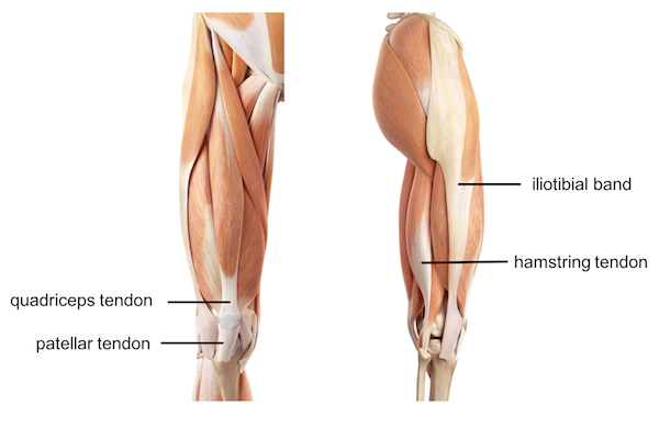
Upper Leg Tendon Anatomy : Muscles of the Leg and Foot - Classic Human Anatomy in ... - Iliotibial band syndrome description the iliotibial band is the tendon attachment of hip muscles into the upper leg (tibia) just below the knee to the outer side of the front of the leg.
Upper Leg Tendon Anatomy : Muscles of the Leg and Foot - Classic Human Anatomy in ... - Iliotibial band syndrome description the iliotibial band is the tendon attachment of hip muscles into the upper leg (tibia) just below the knee to the outer side of the front of the leg.. To download this image, create an account. 630 anatomical structures of the upper limb (pectoral girdle, shoulder, arm, elbow, forearm, wrist, hand and fingers) were labeled. The pads of the machine are situated at the achilles tendon. The achilles tendon (tendo calcaneus or tendo achillis) is the thickest and strongest tendon in the human body. Learn vocabulary, terms and more with flashcards, games and other study tools.
Fibula— a long, thin bone in the lower leg on the lateral side which runs along side the tibia from the knee to the ankle. A tendon is the fibrous tissue that attaches muscle to bone in the human body. Related online courses on physioplus. The tendons of the edl can be palpated on the dorsal surface of the foot. Use the mouse scroll wheel to move the images up and down alternatively use the tiny arrows (>>) on both side of the image to move the images.
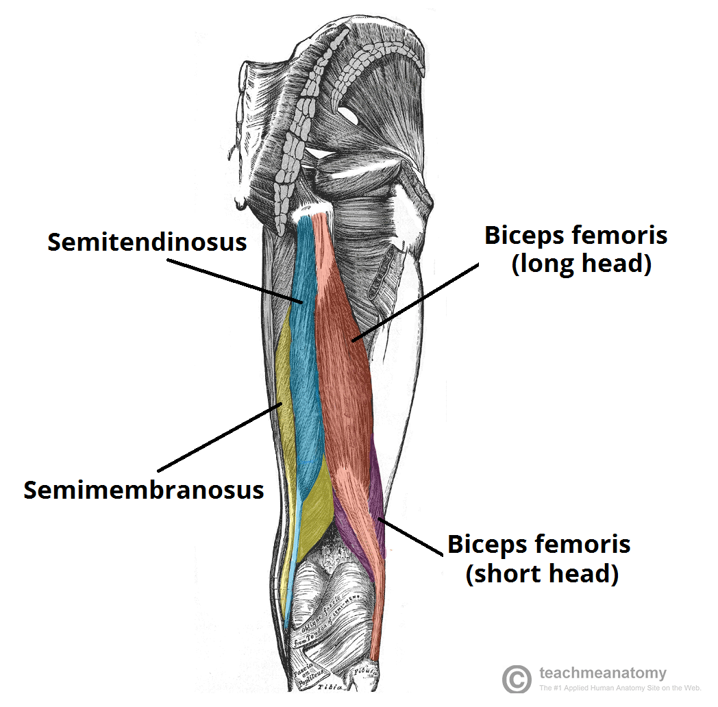
Femur Knee lower leg Anatomy from uploads-ssl.webflow.com The thigh bone, or femur, is the large upper leg bone that connects the lower leg bones (knee joint) to the pelvic bone (hip joint). Superficial veins of upper limb , anatomy : There are four muscles in the anterior compartment of the leg. 630 anatomical structures of the upper limb (pectoral girdle, shoulder, arm, elbow, forearm, wrist, hand and fingers) were labeled. Originates from the lateral condyle of the tibia and the medial surface of the fibula. They're found on the ends of muscles, where they help. The tendons for these muscles begin at your ischial tuberosity, or ischium (the bony bump under each buttock), and attach on the outer edges of your shinbones (tibia and fibula) just below the back of your knee. Learn vocabulary, terms and more with flashcards, games and other study tools.
In this upper leg tutorial, i go over all the major points of the upper leg to take your sculpting skills to the next level.
It's the area that runs from the hip to the knee in each leg. They're found on the ends of muscles, where they help. 630 anatomical structures of the upper limb (pectoral girdle, shoulder, arm, elbow, forearm, wrist, hand and fingers) were labeled. The tendons for these muscles begin at your ischial tuberosity, or ischium (the bony bump under each buttock), and attach on the outer edges of your shinbones (tibia and fibula) just below the back of your knee. Upper limb trauma programme injuries. Fibula— a long, thin bone in the lower leg on the lateral side which runs along side the tibia from the knee to the ankle. ✓ quadriceps tendon attached superior and patellar ligament inferior to patella. Find stockbilleder af concept 3d human upper leg anatomy i hd og millionvis af andre royaltyfri stockbilleder, illustrationer og vektorer i shutterstocks samling. Leg anatomy anatomy poses anatomy study anatomy art anatomy drawing human anatomy anatomy images body reference anatomy anatomical drawings sketchbook ,artist study resources for art students with thanks to artist simone bianchi, how to draw the human figure. Movement at the hip joint occurs when you tendons that help you bend or straighten the knee include: Use the mouse scroll wheel to move the images up and down alternatively use the tiny arrows (>>) on both side of the image to move the images. Collectively, they act to dorsiflex and invert the foot at the ankle joint. Unlike these others, the muscle belly is mostly in the upper part of the.
Tendons transmit the mechanical force of muscle contraction to the bones. There are four muscles in the anterior compartment of the leg. Upper limb trauma programme injuries. Start studying upper leg anatomy. This mri wrist coronal cross sectional anatomy tool is absolutely free to use.
Muscles of the Leg (Human) from anatomycorner.com It attaches the calf muscles to the calcaneus (heelbone) and allows us most of the motion of the ankle is caused by the stronger muscles in the lower leg whose tendons pass by the ankle and connect in the foot. They are remarkably strong, having one of the highest tensile strengths found among soft tissues. Upper legs anatomy — stock image. 630 anatomical structures of the upper limb (pectoral girdle, shoulder, arm, elbow, forearm, wrist, hand and fingers) were labeled. It's the area that runs from the hip to the knee in each leg. The peroneal tendons are in the feet and provide balance and stability during movement. Collectively, they act to dorsiflex and invert the foot at the ankle joint. Want to learn more about it?
Learn vocabulary, terms and more with flashcards, games and other study tools.
What are the functions of patella. Learn about the causes, treatments, and outlook for this condition. See the pictures and anatomy description of knee joint bones, cartilage, ligaments, muscle and tendons with resources for knee problems & injuries. Superficial veins of upper limb , anatomy : 630 anatomical structures of the upper limb (pectoral girdle, shoulder, arm, elbow, forearm, wrist, hand and fingers) were labeled. The lower leg is comprised of two bones, the tibia and the smaller fibula. There are four muscles in the anterior compartment of the leg. Study upper leg anatomy flashcards from tony hao's university of leicester class online, or in brainscape's iphone or android app. Tusindvis af nye billeder af høj kvalitet tilføjes hver dag. Collectively, they act to dorsiflex and invert the foot at the ankle joint. It inserts on the calcaneus. Tendons are fibrous cords attached to muscles and bone. Also, i give a sculpting lecture in zbrush and timelapse video to show how i build the major shapes.
Find stockbilleder af concept 3d human upper leg anatomy i hd og millionvis af andre royaltyfri stockbilleder, illustrationer og vektorer i shutterstocks samling. Tendons are fibrous cords attached to muscles and bone. They have blood vessels and cells to maintain tendon health and repair injured the brachioradialis tendon bends the elbow like the brachialis and biceps. Superficial veins of upper limb , anatomy : Want to learn more about it?
Muscles of the Posterior Thigh - Hamstrings - Damage ... from teachmeanatomy.info Lateral supracondylar line of femur, oblique popliteal ligament of knee insertion: Tendon, tissue that attaches a muscle to other body parts, usually bones. They're found on the ends of muscles, where they help. See the pictures and anatomy description of knee joint bones, cartilage, ligaments, muscle and tendons with resources for knee problems & injuries. The lower leg is comprised of two bones, the tibia and the smaller fibula. ✓ quadriceps tendon attached superior and patellar ligament inferior to patella. Learn about the causes, treatments, and outlook for this condition. Leg anatomy anatomy poses anatomy study anatomy art anatomy drawing human anatomy anatomy images body reference anatomy anatomical drawings sketchbook ,artist study resources for art students with thanks to artist simone bianchi, how to draw the human figure.
To download this image, create an account.
Iliotibial band syndrome description the iliotibial band is the tendon attachment of hip muscles into the upper leg (tibia) just below the knee to the outer side of the front of the leg. To download this image, create an account. See the pictures and anatomy description of knee joint bones, cartilage, ligaments, muscle and tendons with resources for knee problems & injuries. Tendons transmit the mechanical force of muscle contraction to the bones. Upper limb trauma programme injuries. The peroneal tendons are in the feet and provide balance and stability during movement. Posterior surface of calcaneus (via calcaneal tendon). Originates from the lateral condyle of the tibia and the medial surface of the fibula. To describe the mechanical properties of tendons. Tendon, tissue that attaches a muscle to other body parts, usually bones. Want to learn more about it? The posterior upper leg muscles provide your knees with mobility (extension, flexion and. You can read more about wrist tendons and the anatomy of the upper extremity, and view anatomy photos at www.handcare.org.