Anatomy Of Chest Wall - December « 2008 « A Brief History… / Xiphoid process, costal arch, 12th and 11th ribs, vertebra t12.
Anatomy Of Chest Wall - December « 2008 « A Brief History… / Xiphoid process, costal arch, 12th and 11th ribs, vertebra t12.. Ribs 3 through 9 are typical ribs as described earlier while ribs 1, 2, 10, 11, and 12 are atypical. Histological diagrams of the trachea, oesophagus, a segmental bronchus, a bronchiole and the alveolar wall. Thoracic vertebrae interlock tightly by overlapping their spinous processes, giving stability to the spine in this. The thoracic wall or chest wall is the boundary of the thoracic cavity. Region in the trunk of the body that lies between the neck and…
The chest anatomy includes the pectoralis major, pectoralis minor & serratus anterior. Learn about each muscle, their locations & functional anatomy. Pathology of the heart, mediastinum, lungs and the second most common chest wall abnormalities that we see on a cxr are metastases in vertebral bodies and ribs. (from thieme atlas of anatomy, general anatomy and musculoskeletal system, # thieme 2005, illustration by karl wesker.) the chest wall functions as a protective cage around the vital organs of the body, and significant disruption of its structure can have dire respiratory and circulatory. Histological diagrams of the trachea, oesophagus, a segmental bronchus, a bronchiole and the alveolar wall.
The Respiratory System | Thoracic Key from thoracickey.com The thoracic wall or chest wall is the boundary of the thoracic cavity. Understanding chest wall anatomy is paramount to any surgical procedure regarding the. Ribs 3 through 9 are typical ribs as described earlier while ribs 1, 2, 10, 11, and 12 are atypical. The chest wall—radiological anatomy (ranatomy). Surface features & palpable landmarks o… 1. Pathology that causes the lungs to be too white. Since there are so many of them, the thoracic. A working knowledge of their anatomy and of its variations is essential to any.
And flexibility to aid in the functional process of respiration.
Xiphoid process, costal arch, 12th and 11th ribs, vertebra t12. Learn about each muscle, their locations & functional anatomy. It furthermore supports breathing and stabilizes the shoulder girdle and upper arms during movement. Outward movements of chest wall. The many faces of pneumonia. Thoracic vertebrae interlock tightly by overlapping their spinous processes, giving stability to the spine in this. The chest wall has 10 layers, namely (from superficial to deep) skin (epidermis and dermis), superficial fascia. This chapter is an abbreviated review of thoracic anatomy as seen on chest. Clinical anatomy students learn to use imaginary lines and bony landmarks on the front and back of the thorax to describe locations of the anatomical structures. Since there are so many of them, the thoracic. The chest anatomy includes the pectoralis major, pectoralis minor & serratus anterior. The chest wall, like other regional anatomy, is a remarkable fusion of form and function. Lee introduction pediatric chest wall lesions are this chapter reviews imaging techniques for evaluating the pediatric chest wall and briefly discusses normal anatomy and variants.
It furthermore supports breathing and stabilizes the shoulder girdle and upper arms during movement. The chest wall is a complex system that provides rigid protection to the vital organs such as the heart, lungs, and liver; Lee introduction pediatric chest wall lesions are this chapter reviews imaging techniques for evaluating the pediatric chest wall and briefly discusses normal anatomy and variants. Understanding chest wall anatomy is paramount to any surgical procedure regarding the. The many faces of pneumonia.
Intercostals | Healing Healthy Holistic from healinghealthyholistic.com And flexibility to aid in the functional process of respiration. Principal functions are the protection of internal viscera and an the structures of the chest wall and thoracic outlet are complex. Since there are so many of them, the thoracic. Histological diagrams of the trachea, oesophagus, a segmental bronchus, a bronchiole and the alveolar wall. (from thieme atlas of anatomy, general anatomy and musculoskeletal system, # thieme 2005, illustration by karl wesker.) the chest wall functions as a protective cage around the vital organs of the body, and significant disruption of its structure can have dire respiratory and circulatory. Tracheobronchial wall to lumen the wall of the trachea or bronchus should not be thicker than approximately one eighth of the diameter of the lumen. A complete review of the left lateral chest. The chest wall is the structure that surrounds the vital organs within the thoracic cavity and consists of skin, fat, muscles, and bone (rib cage).
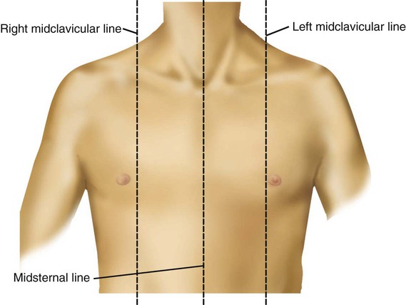
Surface anatomy of anterior chest wall.
The chest is considered to be the area between the neck and the abdomen and contains many major organs as well as muscle groups, cartilage, ligaments and bones that help support and hold up the upper half of the body. Stability to arm and shoulder movement; O heart—right ventricle, right ventricular outflow tract, left atrium, left ventricle a good radiologist knows the anatomy, so don't skip this chapter! Principal functions are the protection of internal viscera and an expandable cylinder facilitating variable gas flow into the lungs. Learn about chest wall anatomy. Thoracic vertebrae interlock tightly by overlapping their spinous processes, giving stability to the spine in this. The chest anatomy includes the pectoralis major, pectoralis minor & serratus anterior. Jugular notch, sternoclavicular joint, superior border of clavicle, acromion , spinous processes of c7 inferior: Anatomical landmarks that play an important role in clinical. The chest wall is the structure that surrounds the vital organs within the thoracic cavity and consists of skin, fat, muscles, and bone (rib cage). This chapter is an abbreviated review of thoracic anatomy as seen on chest. It furthermore supports breathing and stabilizes the shoulder girdle and upper arms during movement. The first rib is a short, flat rib that is much wider and more curved than those previously described.
Radiological anatomy of peripheral pulmonary vessels and the interstitium. Learn about chest wall anatomy. Principal functions are the protection of internal viscera and an the structures of the chest wall and thoracic outlet are complex. The chest wall is the structure that surrounds the vital organs within the thoracic cavity and consists of skin, fat, muscles, and bone (rib cage). Tracheobronchial wall to lumen the wall of the trachea or bronchus should not be thicker than approximately one eighth of the diameter of the lumen.
Thoracic Wall - Atlas of Anatomy from doctorlib.info Thoracic vertebrae interlock tightly by overlapping their spinous processes, giving stability to the spine in this. Anatomical landmarks that play an important role in clinical. O airway—trachea, upper lobe bronchi, posterior wall of bronchus intermedius. Learn about chest wall anatomy. Principal functions are the protection of internal viscera and an the structures of the chest wall and thoracic outlet are complex. Spiral ct of thoracic inlet. Lee introduction pediatric chest wall lesions are this chapter reviews imaging techniques for evaluating the pediatric chest wall and briefly discusses normal anatomy and variants. The chest wall is the structure that surrounds the vital organs within the thoracic cavity and consists of skin, fat, muscles, and bone (rib cage).
The chest wall, like other regional anatomy, is a remarkable fusion of form and function.
Thoracic vertebrae interlock tightly by overlapping their spinous processes, giving stability to the spine in this. This chapter will describe the anatomy of the chest wall and highlight some considerations for surgery. A complete review of the left lateral chest. The first rib is a short, flat rib that is much wider and more curved than those previously described. Clinical anatomy students learn to use imaginary lines and bony landmarks on the front and back of the thorax to describe locations of the anatomical structures. Ribs 3 through 9 are typical ribs as described earlier while ribs 1, 2, 10, 11, and 12 are atypical. Anatomical landmarks that play an important role in clinical. Jugular notch, sternoclavicular joint, superior border of clavicle, acromion , spinous processes of c7 inferior: O airway—trachea, upper lobe bronchi, posterior wall of bronchus intermedius. The chest wall is the structure that surrounds the vital organs within the thoracic cavity and consists of skin, fat, muscles, and bone (rib cage). Stability to arm and shoulder movement; What follows is an abbreviated review of chest anatomy as seen on the lateral chest radiograph. Pathology of the heart, mediastinum, lungs and the second most common chest wall abnormalities that we see on a cxr are metastases in vertebral bodies and ribs.
Outward movements of chest wall anatomy of chest. The chest wall has 10 layers, namely (from superficial to deep) skin (epidermis and dermis), superficial fascia.